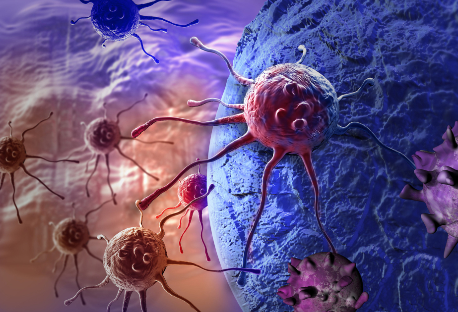
Researchers from Tokyo Metropolitan University have found that the motion of unlabeled cells can be used to tell whether they are cancerous or healthy, according to Phys.org.
They observed malignant fibrosarcoma cells and healthy fibroblasts on a dish and found that tracking and analysis of their paths can be used to differentiate them with up to 94% accuracy.
Beyond diagnosis, their technique may also shed light on cell motility-related functions, like tissue healing. The paper is published in the journal PLOS ONE.
While scientists and medical experts have been looking at cells under the microscope for many centuries, most studies and diagnoses focus on their shape, what they contain, and where different parts are located inside. But cells are dynamic, changing over time, and are known to be able to move.
By accurately tracking and analyzing their motion, we may be able to differentiate cells which have functions relying on cell migration. An important example is cancer metastasis, where the motility of cancerous cells allows them to spread.
However, this is easier said than done. For one, studying a small subset of cells can give biased results. Any accurate diagnostic technique would rely on automated, high-throughput tracking of a significant number of cells.
Many methods then turn to fluorescent labeling, which makes cells much easier to see under the microscope. But this labeling procedure can itself affect their properties. The ultimate goal is an automated method which uses label-free conventional microscopy to characterize cell motility and show whether cells are healthy or not.
Now, a team of researchers from Tokyo Metropolitan University led by Professor Hiromi Miyoshi have come up with a way of tracking cells using phase-contrast microscopy, one of the most common ways of observing cells.
Phase-contrast microscopy is entirely label-free, allowing cells to move about on a petri dish closer to their native state, and is not affected by the optical properties of the plastic petri dishes through which cells are imaged.
Through innovative image analysis, they were able to extract the trajectories of many individual cells. They focused on properties of the paths taken, like migration speed, and how curvy the paths were, all of which would encode subtle differences in deformation and movement.
As a test, they compared healthy fibroblast cells, the key component of animal tissue, and malignant fibrosarcoma cells, cancerous cells which derive from fibrous connective tissue. They were able to show that the cells migrated in subtly different ways, as characterized by the "sum of turn angles" (how curvy the paths were), the frequency of shallow turns, and how quickly they moved.
In fact, by combining both the sum of turn angles and how often they made shallow turns, they could predict whether a cell was cancerous or not with an accuracy of 94%.
The team's work not only promises a new way to discriminate cancer cells, but applications to research of any biological function based on cell motility, like the healing of wounds and tissue growth.